Shoulder Tendon And Ligament Anatomy : Posterior Tibiofibular Ligament - Earth's Lab - Ligaments tendons and other stuff.. The achilles tendon connects the heel to the calf muscle and is essential for running, jumping, and standing on the toes. Anatomy is the amazing science. Injury of tendons and ligaments remodel with scar formation with differences in themselves. Tendons and ligaments are bands of connective tissue that help stabilize the body and allow movement. Learn about the muscles, tendons, bones, and ligaments that comprise the knee joint anatomy.
Anteriorly the subscapularis tendon is separated from the supraspinatus tendon by a gap, the rotator interval another important ligament, the coracoacromial ligament (cal). Brachial plexus, axillary artery, brachiocephalic vein, axillary region. Once stretched, they tend to stay. The shoulder joint is the articulation between the glenoid cavity of the scapula and the head of the humerus. Links the coracoid to the acromium and forms the.
Shoulder Sergery | Shoulder anatomy, Shoulder joint ... from i.pinimg.com In order to achieve this flexibility but maintain a stable shoulder, there is a complex interplay between the joints, muscles and ligaments. This instability is countered by the strength of the rotator cuff muscles, tendons, ligaments, and the glenoid labrum. Other smaller muscles and tendons surround the knee joint as well. The achilles tendon connects the heel to the calf muscle and is essential for running, jumping, and standing on the toes. Ligaments tendons and other stuff. Contents of ri = long head of biceps tendon, superior glenohumeral ligament, glenohumeral capsule. Anatomy is the amazing science. There are several important ligaments in the shoulder.
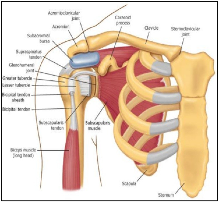
The shoulder is comprised of a ball (humerus) and socket (scapula), bones, ligaments, tendons and muscles that move the arms and connect them to the torso.
Tendons and ligaments are bands of connective tissue that help stabilize the body and allow movement. Tendon and ligament injuries often go hand in hand with horses involved in vigorous athletic pursuits. Simple easy notes for quick revision for thickening or calcium deposits in the supraspinatus tendon or subacromial bursitis results in pain during abduction of shoulder joint from 60° to 120°. This is the currently selected item. Synovial fluid is constantly, but slowly being replenished think of the way water in a pool is not typically drained completely but is gradually replaced. Once stretched, they tend to stay. Ligaments are soft tissue structures that connect bones to bones. The shoulder is comprised of a ball (humerus) and socket (scapula), bones, ligaments, tendons and muscles that move the arms and connect them to the torso. Anteriorly the subscapularis tendon is separated from the supraspinatus tendon by a gap, the rotator interval another important ligament, the coracoacromial ligament (cal). Start studying shoulder ligaments and tendons. It can help you understand our world more detailed and specific. The accessory ligaments for shoulder are coracohumeral ligament, coracoacromial ligament, glenohumeral ligament, and transverse humeral ligament and they are part of the rotator interval, which a space between the subscapularis tendon and the supraspinatus tendon. The achilles tendon connects the heel to the calf muscle and is essential for running, jumping, and standing on the toes.
Transverse humeral ligament (thl) :holds the tendon of the long head of biceps brachii muscle in the groove between the greater and lesser tubercle on the humerus (intertubercular sulcus). Muscles allow us to move by pulling on bones. Brachial plexus, axillary artery, brachiocephalic vein, axillary region. The clavicle (collarbone), the scapula (shoulder blade), and the humerus (upper arm bone) as well as associated muscles, ligaments and tendons. Anatomy of the shoulder joint (scapula, humerus, glenoid labrum, tendons) of the dog on ct.
Repetitive H-Wave® device stimulation and program induces ... from media.springernature.com Simple easy notes for quick revision for thickening or calcium deposits in the supraspinatus tendon or subacromial bursitis results in pain during abduction of shoulder joint from 60° to 120°. Learn about the muscles, tendons, bones, and ligaments that comprise the knee joint anatomy. The patellar tendon on the front of the knee is part of the quadriceps mechanism. Anatomy of the human body via wikimedia commons, public domain. Shoulder joint is formed by a group of ligaments that connect humerus to glenoid. Tendons and ligaments are bands of connective tissue that help stabilize the body and allow movement. Related posts of shoulder muscles tendons anatomy. Other smaller muscles and tendons surround the knee joint as well.
In order to achieve this flexibility but maintain a stable shoulder, there is a complex interplay between the joints, muscles and ligaments.
Superior glenohumeral ligament and coracohumeral ligament are the primary restraints to posterior acromioclavicular ligament anatomy. The human shoulder is made up of three bones: In addition to the bones and joints, the shoulder contains a network of soft tissues, such as muscles, tendons, and ligaments. Ac joint is a diathrodial joint with a fibrocartilaginous disk. Transverse humeral ligament (thl) :holds the tendon of the long head of biceps brachii muscle in the groove between the greater and lesser tubercle on the humerus (intertubercular sulcus). Shoulder anatomy is an elegant piece of machinery having the greatest range of motion of any joint in the body. Although scarring depends on the quality and quantity of the injured tissues, it can be. The bones of the shoulder consist of the humerus (upper arm bone), the scapula. Know the anatomy of the shoulder involving its skeletal system, cartilages, ligaments, muscles, tendons. The shoulder joint is the articulation between the glenoid cavity of the scapula and the head of the humerus. Anatomy of the shoulder joint (scapula, humerus, glenoid labrum, tendons) of the dog on ct. Tendons and ligaments are complex structures and have different anatomical and dynamic properties. Brachial plexus, axillary artery, brachiocephalic vein, axillary region.
Ligaments aid in joint stability during rest and movement and help prevent injury from hyperextension and hyperflexion (excessive movements). It can help you understand our world more detailed and specific. There are several important ligaments in the shoulder. Ligaments and tendons are fibrous bands of connective tissue that attach to bone connecting two or more bones together and help stabilize joints. Other smaller muscles and tendons surround the knee joint as well.
Anatomy Block I - Back and Shoulder at Rice University ... from classconnection.s3.amazonaws.com Although scarring depends on the quality and quantity of the injured tissues, it can be. The shoulder is comprised of a ball (humerus) and socket (scapula), bones, ligaments, tendons and muscles that move the arms and connect them to the torso. Anteriorly the subscapularis tendon is separated from the supraspinatus tendon by a gap, the rotator interval another important ligament, the coracoacromial ligament (cal). For more anatomy content please follow us and visit our website: Tendons and ligaments are complex structures and have different anatomical and dynamic properties. A joint capsule is a watertight sac that surrounds a joint. The accessory ligaments for shoulder are coracohumeral ligament, coracoacromial ligament, glenohumeral ligament, and transverse humeral ligament and they are part of the rotator interval, which a space between the subscapularis tendon and the supraspinatus tendon. Anatomy of the human body via wikimedia commons, public domain.
Although scarring depends on the quality and quantity of the injured tissues, it can be.
In addition to the bones and joints, the shoulder contains a network of soft tissues, such as muscles, tendons, and ligaments. In the horse, lateral and medial movements of this joint are impossible due to the shape of the humeral head; Simple easy notes for quick revision for thickening or calcium deposits in the supraspinatus tendon or subacromial bursitis results in pain during abduction of shoulder joint from 60° to 120°. The shoulder is comprised of a ball (humerus) and socket (scapula), bones, ligaments, tendons and muscles that move the arms and connect them to the torso. This is the currently selected item. Superior glenohumeral ligament and coracohumeral ligament are the primary restraints to posterior acromioclavicular ligament anatomy. For more anatomy content please follow us and visit our website: Once stretched, they tend to stay. Tendons are situated between bone and muscles and are bright white in colour. Related posts of shoulder muscles tendons anatomy. Anteriorly the subscapularis tendon is separated from the supraspinatus tendon by a gap, the rotator interval another important ligament, the coracoacromial ligament (cal). It can help you understand our world more detailed and specific. Movement is therefore limited to flexion and extension.
The patellar tendon on the front of the knee is part of the quadriceps mechanism shoulder tendon anatomy. Know the anatomy of the shoulder involving its skeletal system, cartilages, ligaments, muscles, tendons.


0 Comments